

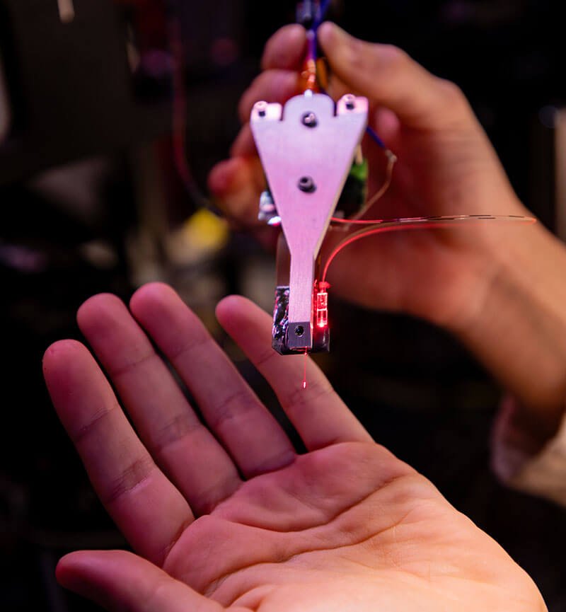
Neuropixels probes are allowing scientists to track the electrical activity of hundreds of individual brain cells at once. Researchers say the instruments are reshaping experiments on movement, perception and decision-making, while early human surgical studies show both their promise and their limitations.
A November 2, 2026 technology feature in Nature documents how Neuropixels, developed through a collaboration that includes the Howard Hughes Medical Institute’s Janelia Research Campus in Virginia and imec in Belgium, has progressed from animal experiments to specialized recordings during human brain surgery.
Neuropixels is a research instrument, not a device that reads a person’s thoughts or a proven treatment for brain disease. Its breakthrough is in measurement: recording signals from many individual neurons at high speed and across multiple brain regions.
What makes a Neuropixels probe different?
Each probe is a narrow silicon shaft covered with microscopic electrical recording sites. Those sensors detect voltage changes produced by nearby active neurons. Electronics on the device amplify and digitize the signals, while software works to distinguish individual neurons from the combined recordings.
The original Neuropixels 1.0 design, described in a 2017 Nature paper, featured 960 recording sites, with 384 channels read simultaneously. The Neuropixels 2.0 four-shank design has 5,120 recording sites and can read 1,536 channels at once, according to the 2021 Science paper. A channel is not automatically one neuron: researchers must use spike-sorting and quality-control methods to identify distinct units.
The technology provides finer temporal detail than methods that measure averaged electrical signals across large groups of cells. That precision helps scientists investigate how neurons respond together when an animal sees, moves or makes a choice.
A brain-wide project recorded more than 620,000 neuronal units
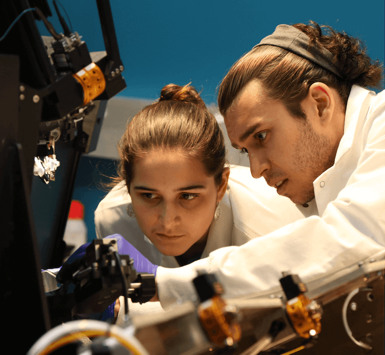
In September 2025, the International Brain Laboratory published a standardized, multicenter mouse study involving 12 laboratories, 139 mice and 699 Neuropixels probe insertions. The team collected 621,733 recorded units across 279 brain areas while the animals performed tasks combining sensory input, movement and decisions. The peer-reviewed study emphasizes the importance of comparing recordings made with common protocols.
There is an important qualification: the project’s data documentation reports that 75,708 units met its good-quality criteria. The total recording count should not be misrepresented as an equal number of perfectly isolated, validated cells.
Such projects are changing the field’s understanding of brain organization. Neural signals linked to seeing, moving and choosing can be distributed across multiple regions rather than confined neatly to single locations.
Human neurosurgery offers a limited but revealing window
A 2022 Nature Neuroscience study demonstrated recordings from more than 200 well-isolated single units in human cerebral cortex during neurosurgical procedures. Unlike routine scalp-based tests, the method involves placing a probe directly into brain tissue, making it possible only under tightly controlled research and surgical circumstances.
In May 2026, investigators reported recordings in seven patients under general anesthesia during temporal-lobe surgery. The study in Nature identified 651 neural units and found that some hippocampal activity continued to distinguish sounds and respond to features of spoken language. The finding suggests complex neural processing can persist under anesthesia; it does not prove that the patients experienced conscious awareness of the sounds.
Other research has focused on epilepsy. A 2026 study in Nature Neuroscience examined 1,152 neurons during 1,094 interictal epileptiform discharges at nine human cortical sites. Researchers found activity patterns that sometimes preceded these between-seizure discharges by up to one second. This was a research finding about a specific electrical event, not a validated system for predicting every clinical seizure or evidence of a new approved therapy.
Neuropixels Opto adds light-based control
Researchers are now extending the probes beyond passive recording. In a June 2026 Nature Methods study, teams at the Allen Institute, University of Washington, UCL, Janelia and imec described a prototype Neuropixels Opto probe. It combines 960 recording sites with 28 light emitters on a silicon shaft approximately 70 micrometers wide and one centimeter long.
In experiments with genetically prepared mice, light pulses could activate or suppress selected groups of neurons while nearby activity was recorded. This approach lets scientists test potential causal roles of particular cell groups instead of relying only on correlations between activity and behavior. The device remains a research prototype; the published experiments do not establish a treatment for patients.
Why this matters in the United States
American institutions—including the Allen Institute in Seattle, Janelia in Virginia, the University of Washington and Massachusetts General Hospital—have played significant roles in the research. The National Institutes of Health’s BRAIN Initiative has also supported development of advanced neural recording tools.
More detailed measurements may eventually help scientists understand why circuits malfunction in epilepsy, movement disorders and some neuropsychiatric conditions. But those possibilities require years of further validation. Neuropixels probes are invasive, can sample only a fraction of the brain, and generate large datasets that require extensive interpretation. Human use requires careful attention to consent, surgical risk, data privacy and whether discoveries from animal models generalize to patients.
What comes next
The next challenge is not simply recording more neurons. Scientists need to demonstrate which recordings are reliable across time, how neural populations interact, and whether a finding can lead to safe, effective clinical intervention. The most consequential outcome may be a more rigorous understanding of brain circuits—not a machine that can decode every thought.
Video: Neuropixels data explained
The Allen Institute’s official Neuropixels Dataset Tutorial (June 13, 2024) explains the mouse visual-system dataset and how researchers analyze its electrical recordings.
Primary sources and further reading
- Nature technology feature, Diana Kwon, November 2, 2026
- International Brain Laboratory, brain-wide activity map, Nature, 2025
- Paulk and colleagues, intraoperative human recordings, Nature Neuroscience, 2022
- Katlowitz and colleagues, hippocampal activity under anesthesia, Nature, 2026
- Silva and colleagues, epileptiform discharges, Nature Neuroscience, 2026
- Lakunina and colleagues, Neuropixels Opto, Nature Methods, 2026
- Allen Institute explanation and original documentary images, June 2026