


LA JOLLA, Calif. — Scientists at the Salk Institute for Biological Studies have mapped 1,067 previously overlooked microproteins in the human frontal cortex, finding that some change in abundance in brain tissue from people with Alzheimer’s disease. Their research opens questions about the brain’s molecular machinery but does not establish a new treatment or cure.
The peer-reviewed study was published in Nature Aging on September 14, 2026. The authors include researchers from Salk in California and Rush University Medical Center in Illinois. It attracted renewed coverage from PsyPost on November 3.
What are microproteins, and why were they overlooked?
Microproteins are short proteins made from compact stretches of genetic instructions, often consisting of no more than 150 amino acids. Traditional reference databases and detection methods tend to focus on larger, more familiar proteins. Small protein-producing sequences may also overlap known genes, making their individual contributions difficult to distinguish.
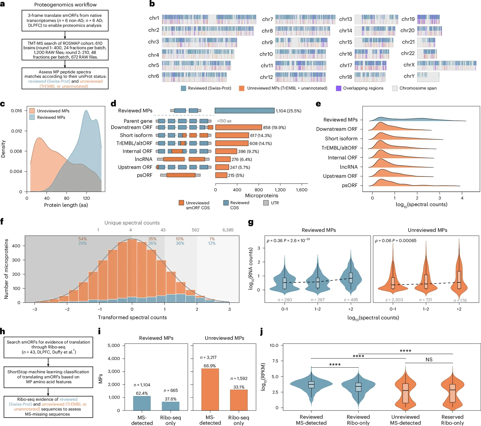
The researchers assembled a custom search database from RNA data collected from 12 donated brains. They then analyzed mass-spectrometry data from 610 postmortem dorsolateral prefrontal-cortex samples in the Religious Orders Study and Rush Memory and Aging Project. Their broad search identified 4,321 short proteins, including both previously cataloged and unreviewed sequences. A more demanding evaluation, using experimentally observed protein fragments and computer-predicted spectra, identified 1,067 unreviewed microproteins with strong evidence.
This atlas is a map of the frontal cortex, not a complete inventory of every region of the brain. Nor does the presence of a microprotein establish its role in disease.
The microprotein linked to cell energy
One especially interesting finding involved micro-MKKS63, a 63-amino-acid protein generated from the MKKS genetic region. In the frontal-cortex measurements, it was detected much more prominently than the larger conventional MKKS protein. Its abundance was lower in samples from people with symptomatic Alzheimer’s disease.
The researchers tested its function in cultured human microglia, immune cells that help protect and maintain brain tissue. After selectively disrupting the DNA sequence responsible for micro-MKKS63, they measured reduced mitochondrial oxygen consumption and energy-related activity. This supports a role for the microprotein in microglial energy metabolism.
However, these were laboratory-cell experiments. They do not show that a shortage of micro-MKKS63 causes Alzheimer’s disease in humans, nor that increasing it would improve memory or reverse neurodegeneration.
New clues about how genes work
The investigators found that some microproteins change separately from the better-known protein products associated with the same genes. This suggests that measuring only a gene’s primary protein may miss biologically relevant changes. They also reported evidence of small proteins generated from some pseudogenes, DNA sequences sometimes regarded as inactive remnants.
For another candidate linked to ACTBP7, experimental expression changed the arrangement of structural fibers in cultured microglia. These findings provide starting points for more targeted experiments, not established explanations of Alzheimer’s.
What the findings mean for American families
Alzheimer’s disease affects families, care systems and researchers throughout the United States. The immediate importance of this study is the publicly accessible research resource, rather than any change in clinical care. Other laboratories can examine the brain microprotein atlas, investigate specific molecules and test findings in additional tissue and experimental systems.
Because the brain specimens were collected after death, differences between Alzheimer’s and non-Alzheimer’s groups cannot by themselves establish cause and effect. Microproteins may be contributors to disease, responses to damage, or markers of other cellular changes. Additional research is required to distinguish those possibilities.
Sources and further reading
- Original open-access study in Nature Aging (September 14, 2026; DOI 10.1038/s43587-026-01207-x).
- Salk Institute research announcement (September 14, 2026).
- Independent coverage in Nature (September 16, 2026).
- PsyPost reporting (November 3, 2026).
Image note: Featured image (Figure 7) and inline Figure 1 reproduce original research figures from Miller et al., Nature Aging (2026), distributed under CC BY 4.0. Neither image depicts a patient or clinical treatment. This study does not demonstrate a clinical therapy.